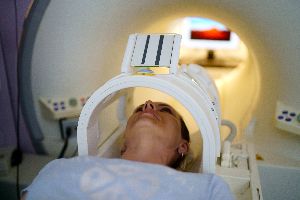
Medical imaging is a broad term for an array of tools that use high-energy waves to produce images of the inside of your body. They allow doctors to spot injuries or illnesses that can't be seen with the naked eye. Each type is used for a specific purpose, so whichever you come across depends on your symptoms. Here are the most common ones used to diagnose and treat patients.
A Guide to Standard Medical Imaging Techniques
1. X-Rays
X-rays use light radiation to obtain images of a patient's bones, organs, and blood vessels. They can help diagnose any problems a doctor is looking for, from fractures or infections to diseases like pneumonia or breast cancer. Because they can detect various issues and provide a deeper understanding of what their patients are experiencing, they are commonly used in hospitals and private clinics.
2. CT Scans
A doctor can obtain a series of scans using special X-Ray equipment, along with computers and sophisticated software. The images get compiled into a detailed 3D representation, providing a level of detail that would otherwise take several X-Rays to achieve. This makes a CT scan more cost-effective and faster than a traditional method, helping to diagnose conditions quickly and efficiently.
3. MRI Scans
This type of medical imaging uses an extremely powerful nonionizing magnet that applies a strong magnetic force to organs and tissues to generate a scan. It also utilizes low-energy radio waves and computer algorithms to refine and clarify the resulting image.
This scan provides significant insight into how certain organs are functioning and any abnormalities. Doctors often use it to examine the brain, spine, and joints.
4. Ultrasounds

An ultrasound uses high-frequency sound waves to create images. While it's most often used to look at a developing baby during pregnancy, it can also be used to diagnose and evaluate other conditions. Common applications include diagnosing diseases in the abdominal organs, detecting problems in veins and arteries, or looking for issues in the musculoskeletal system.
5. PET Scans
A positron emission tomography (PET) scan detects and marks levels of radioactivity within organs or other tissues. Typically, it takes about 20 to 30 minutes to complete. It uses a radioactive drug—a tracer—to track and provide details on specific bodily processes, with the scanner reading the radiation given off. The imaging can be used to map out any changes in metabolism associated with the areas in question.
For reliable and efficient medical imaging, visit Independence Healthcare in Soldotna, AK. Their doctors, nurse practitioners, and physician assistants take pride in helping their patients become as informed as possible. From orthopedics to sports medicine, they offer many services to keep you healthy. Visit their website to learn more about their treatment options, and call (907) 262-6454 to schedule an appointment today.
About the Business
Have a question? Ask the experts!
Send your question

